
Ankle and foot examination include objectives, Assessment, Diagnose, and Treat. ankle joint is a synovial joint of the lower limb. it transfers whole body weight to the ground
 |
| Examination of the Foot and Ankle |
- The ankle and foot is a complex structure comprised of 28 bones (including 2 sesamoid bones) and 55 articulations (including 30 synovial joints), interconnected by ligaments and muscles.
- In addition to sustaining substantial forces, the foot and ankle serve to convert the rotational movements that occur with weight-bearing activities into sagittal, frontal, and transverse movements.

- The ankle joint (or talocrural joint) is a synovial joint located in the lower limb. It is formed by the bones of the leg (tibia and fibula) and the foot (talus).
- Functionally, it is a hinge type joint, permitting dorsiflexion and plantarflexion of the foot.
Articulating Surfaces
- The ankle joint is formed by three bones; the tibia and fibula of the leg, and the talus of the foot:
- The tibia and fibula are bound together by strong tibiofibular ligaments. Together, they form a bracket shaped socket, covered in hyaline cartilage. This socket is known as a mortise.
- The body of the talus fits snugly into the mortise formed by the bones of the leg. The articulating part of the talus is wedge-shaped – it is broad anteriorly, and narrow posteriorly:
- Dorsiflexion – the anterior part of the talus is held in the mortise, and the joint is more stable.
- Plantarflexion – the posterior part of the talus is held in the mortise, and the joint is less stable.
Ligaments
- There are two main sets of ligaments, which originate from each malleolus.
- Medial Ligament: The medial ligament (or deltoid ligament) is attached to the medial malleolus (a bony prominence projecting from the medial aspect of the distal tibia).
- It consists of four ligaments, which fan out from the malleolus, attaching to the talus, calcaneus, and navicular bones. The primary action of the medial ligament is to resist over-eversion of the foot.
- Lateral Ligament: The lateral ligament originates from the lateral malleolus (a bony prominence projecting from the lateral aspect of the distal fibula).
- It resists over-inversion of the foot, and is comprised of three distinct and separate ligaments:
- Anterior talofibular Ligament – spans between the lateral malleolus and lateral aspect of the talus.
- Posterior talofibular Ligament – spans between the lateral malleolus and the posterior aspect of the talus.
Movements and Muscles Involved
- The ankle joint is a hinge type joint, with movement permitted in one plane.
- Thus, plantarflexion and dorsiflexion are the main movements that occur at the ankle joint. Eversion and inversion are produced at the other joints of the foot, such as the subtalar joint.
- Plantarflexion – produced by the muscles in the posterior compartment of the leg (gastrocnemius, soleus, plantaris, and posterior tibialis).
- Dorsiflexion – produced by the muscles in the anterior compartment of the leg (tibialis anterior, extensor hallucis longus, and extensor digitorum longus).
History for Ankle and foot examination
- Take a HISTORY
- What is the patient’s chief complaint?
- Pain?
- Where?
- When?
- How bad?
- What is it like?
- What makes it better?
- What makes it worse?
- Acute Injury vs. Chronic –?
- Progression of Symptoms?
- pain scale -visual analog scale
Background Information
- Any Previous Injuries
- Past Surgical History
- Past Medical History
- Medications
- Allergies
- Social History – Work situation (laboring type job?) Home situation
Observation in Ankle and foot examination
- Body Built
- Posture: anterior, lateral and posterior view
- Weight-bearing: equal on both sides Compare weight-bearing and non-weight-bearing position of the foot in
- See for Contour of Foot soft tissue swelling Bony callosity
Deformities
Forefoot Varus
- midtarsal joint- Inversion
- Subtalar joint- Neutral
- Forefoot – Valgus
- midtarsal joint -Eversion
- Subtalar joint- Neutral.
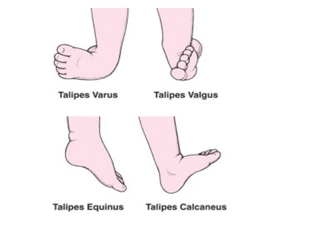
- Talipes Eqinus: Plantar flexed foot Can cause plantar fascitis, metatarsalgia.
- Claw Toes: MTP joint in Hyper Extension, IP joint in Flexion
- Hammer Toes: MTP in Hyperextended, PIP joints are Flexed and DIP are Hyperextended
- Hallux Rigidus: Stiffness of Great toe at MTP which Maybe due to OA
- Splay foot : Spread of Metatarsal
- Rocker Bottom Foo:t Forefoot in dorsiflexion and longtudinal Arch may be absent
Standing and Weight-bearing:
- Anteroposterior view
- Weight Bearing: Equal on both feet and forefoot/hindfoot?
- Position of foot Supination/pronation ?
- Ask the Patient to walk on heel and toes: Gives the idea about muscle power or functional range of motion • Does the patient use Cane or stick? Use of cane on the opposite side decrease the load on the ankle by 1/3 of body weight
- Check the toes if parallel/ straight/
- check for Spurs/ exostosis/Swelling
- Check for tibia/ knee
- Lateral view
- Observe the longitudinal arch of the foot
- The medial longitudinal arch should be higher than the lateral
- Posterior view
- measure Bulk of calf: compare on both sides
- check Achilles tendon: Vertical on both sides
- Observe calcaneum for shape position callosity
- check the Position of the malleolus
- Foot Print Pattern
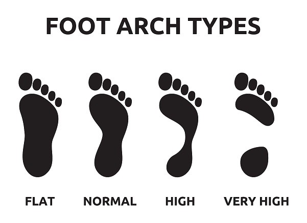
- it will provide information about arches of the foot
- stride length
- step length
- use Light film of baby’s oil on patient foot and apply powder
- Ask the patient to step on a piece of colored paper
- Observe for a pattern of foot
Palpation Ankle and foot examination
- SURFACE ANATOMY IS THE KEY!!!
- Palpate for the local rise of temperature Local tenderness Palpation of specific areas
- Palpation(Bony) : Medial aspect
- Palpation(Bony) : Lateral Aspect
- Palpation (soft tissue):
- Zone 1: Head of 1st MT bone: Pathology – gout, hallux valgus
- Zone 2: Navicular tubercle and talar head
- Zone 3: Medial malleolus Palpate the Deltoid ligament, palpate the following structure in the depression between the posterior aspect of medial malleoli and Achilles tendon Tibialis posterior tendon, Flexor digitorum longus tendon, Posterior tibial artery and tibial nerve, Flexor hallucis longus tendon
- Zone 4: Dorsum of the foot between malleoli there are 3 important tendons and one vessel that passes between the malleoli. From medial to lateral they are: Tibialis anterior tendon Extensor hallucis longus tendon, Dorsal pedal artery, Extensor digitorum longus tendon, Peroneus Tertius
- Zone 5: Lateral Malleoli
- clinically important ligaments, which comprise the lateral collateral ligaments of the ankle joint. From anterior to posterior, they are: -Anterior talofibular ligament
- Calcaneofibular ligament
- Posterior talofibular ligament
- Zone 6:sinus tarsi commonly involved in an ankle sprain
- Zone 7:head of 5th MT Tailors bunion
- Zone 8:Calcaneum Retrocalcaneal bursa/ calcaneal bursa
- Zone 9: plantar surface
Examination in Ankle and foot examination
- Examination of the foot and ankle STEPS in the PHYSICAL EXAMINATION Consent Privacy Exposure Gait analysis Observation Palpation Range of motion Neurovascular assessment Special tests
- Exposure Both shoes and socks off. At least have trousers rolled up to the knees, preferably down to underwear
Gait Analysis
- OBJECTIVES
- Identify the phases of gait and perform a functional gait analysis.
- GAIT ANALYSIS STRIDE LENGTH •
- Symmetrical side-to-side?
- Shortened? FOOT PROGRESSION
- Symmetrical?
- Neutral?
- Internal?
- External?
Range of Motion
Range of Motion Ankle motion Check the range of motion

- Dorsiflexion- 10 to 30 -Reduce the talonavicular joint
- Plantar flexion – 20 to 50
- Inversion and Eversion: Patient sitting on a stool with the knee flexed at 70 degrees Hold ankle firmly from dorsum to fix talus by dorsiflexion, Hold the body of calcaneum in between thumb on one side and index and middle finger on another side with another hand now Turn in for inversion and turn out for eversion • I= 35-degree E= 25 degree
- Range of Motion Adduction and Abduction of Forefoot
- Hold hindfoot from dorsum with one hand Hold forefoot with other hand and Passively deviate forefoot inward for adduction and outward for adduction
- First MTP joint motion this joint is Principally involved in toe-off phase of gait • Stabilize foot and move great toe through flexion and extension
NEUROVASCULAR ASSESSMENT
Motor
- Dorsiflexion: Tibialis Anterior Deep Peroneal Nerve L4
- Extensor Hallucis Longus L5
- Extensor Digitorum Longus L5
- Plantar Flexors : Peroneus Longus and Brevis
- Superficial peroneal Nerve, S1
- Gastrnemius and Soleus – Tibial Nerve, S1 S2
- Flexor Digitorum Longus -Tibial Nerve L5
- Tibialis Posterior – Tibial Nerve L5
- Ankle Reflex: indicates the integrity of s1
Special test in Ankle and foot examination
Stress test For medial and lateral collateral ligament
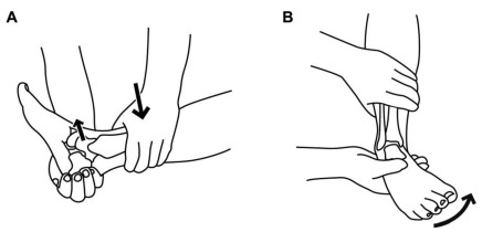
- Place the ankle in neutral position Hold the lower leg firmly from the front by one hand Hold the foot at about the level of talus by opposite hand For testing the lateral collateral ligament, invert the foot and for testing of medial collateral ligament stress has to be given in opposite direction.
- Anterior Drawer test : For the integrity of capsule and anterior talofibular ligament Pulling the heel anteromedially against resistance applied by the other hand over the anterior aspect of lower leg Anterior subluxation of 3 mm of the talus is pathological
- Test for rupture of tendon-Achilles
- Thompson test: Prone position with feet projecting beyond examining table Calf muscle squeezed
- Normal or partially torn- plantar flexion
- Complete rupture- No movement of the foot
- Test for pre-Achilles and post Achilles pathologies
- Pt asked to walk on toes with the heel off the ground pain in pre-Achilles pathology
- Walk on heel– pain in post-Achilles pathology
- Achilles tendonitis – pain in both mode of walking, more on walking on toes
- Ankle Dorsiflexion Test:To determine whether gastrocnemius or soleus causing limitation of ankle dorsiflexion With flexion of the knee joint, ankle dorsiflexion achieved – Gastronemius Not affected by flexion of the knee- Soleus
- Homan’s sign: Test for deep vein thrombophlebitis
- Forcibly dorsiflex ankle with a leg in extension causes Pain in the calf muscle
- Measurement of equinus deformity
- Position- lying on the bed on lateral position Passively dorsiflex as far as possible Measure angle between the long axis of the leg and the long axis of midfoot Subtract 90 from an angle
- Tibial Torsion Test: To determine whether toeing in is due to internal rotation of tibia Normally a line drawn between malleoli is rotated externally 15 degrees from a perpendicular line drawn from the tibial tubercle to the ankle In tibial torsion, the malleolar line may face directly anteriorly close to the perpendicular line.
- Forefoot Adduction Correction Test: Forefoot adduction is common in children which may or may not need correction If adduction can be corrected manually and abduction can be done beyond neutral position – NO TREATMENT If only partially corrected to neutral or less than neutral – CAST CORRECTION
- Colman Block test – Coleman block test evaluates hindfoot flexibility and pronation of forefoot; initial deformity is in the forefoot followed by subsequent changes in the hindfoot test is performed by placing the patient’s foot on a woodblock, 2.5 to 4 cm thick, with the heel and lateral border of the foot on the block and bearing full weight while the first, second, & 3rd metatarsals are allowed to hang freely into plantar flexion and pronation.
- Interpretation: – if heel varus corrects while the patient is standing on the block, hindfoot is considered flexible if the subtalar joint is supple & correct w/ block test, then surgical procedures may be directed to correcting forefoot pronation, which is usually due to plantar flexion of 1st metatarsal
- if hindfoot is rigid, then surgical correction of both forefoot & hindfoot are required
- Examination of footwear
- Distortion of shape: underlying rigid deformity
- Wrinkling of footwear- in persistent varus of the heel, deep wrinkles on the inner aspect of the heel
- Bulging out thinning
- Deformity of sole
0Comments