
The Brainstem lies at the base of the brain and the top of the spinal cord.
The brainstem is the structure that connects the cerebrum of the brain to the spinal cord and cerebellum.
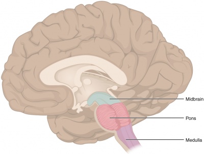
- It is composed of 3 sections in descending order: the midbrain, pons, and medulla oblongata.
- It is responsible for many vital functions of life, such as breathing, consciousness, blood pressure, heart rate, and sleep.
The brainstem contains many critical collections of white and grey matter.
 |
| Brainstem |
- The grey matter within the brainstem consists of nerve cell bodies and form many important brainstem nuclei. Ten of the twelve cranial nerves arise from their cranial nerve nuclei in the brainstem
- The white matter tracts of the brainstem include axons of nerves traversing their course to different structures; the axons originate from cell bodies located elsewhere within the central nervous system (CNS). Some of the white matter tract cell bodies are located within the brainstem as well. These tracts travel both to the brain (afferent) and from the brain (efferent) such as the somatosensory pathways and the corticospinal tracts, respectively.
Although it is the most evolutionarily ancient part of our brain, the brainstem is still very complex and important.
- The brainstem may not provide us with the higher intelligence we normally associate with being human, but it does carry all of the information to and from those areas we do associate with higher intelligence.
- It ensures the vital functions necessary to support those areas continue uninterrupted.
Structure
 |
| Brainstem |
The brainstem is generally said to be composed of three parts.
Components, from above downward:
- Midbrain (or Mesencephalon)
- Pons (part of the metencephalon)
- Medulla AKA Medulla Oblongata (myelencephalon)
Midbrain
- The upper posterior (i.e. rear) portion of the midbrain is called the tectum, which means "roof." The surface of the tectum is covered with four bumps representing two paired structures: the superior and inferior colliculi. The superior colliculi are involved in eye movements and visual processing, while the inferior colliculi are involved in auditory processing.
- Another important nucleus,the substantia nigra, is located here. The substantia nigra is rich in dopamine neurons and is considered part of the basal ganglia. In Parkinson's disease, neurodegeneration occurs in the substantia nigra, and this neurodegeneration is associated with the hallmark movement dysfunction we see in Parkinson's.
Pons
- An important pathway for tracts that run from the cerebrum down to the medulla and spinal cord, as well as for tracts that travel up into the brain. It also forms important connections with the cerebellum via fiber bundles known as the cerebellar peduncles.
- Home to a number of nuclei for cranial nerves.
- Nerves that carry information about sensations of touch, pain, and temperature from the face and head synapse in a nucleus in the pons.
- Motor commands dealing with eye movement, chewing, and facial expressions also originate in the pons.
- Additionally, cranial nerve nuclei in the pons are involved in a number of other functions, including swallowing, tear production, hearing, and maintaining balance/equilibrium.
Medulla
- The point where the brainstem connects to the spinal cord
- Contains a nucleus called the nucleus of the solitary tract that is crucial for our survival (receives information about blood flow, along with information about levels of oxygen and carbon dioxide in the blood, from the heart and major blood vessels). When this information suggests a discordance with bodily needs (e.g. blood pressure is too low), there are reflexive actions initiated in the nucleus of the solitary tract to bring things back to within the desired range.
- Essential to our survival because it ensures vital systems eg cardiovascular and respiratory systems are working properly.
- Responsible for a number of reflexive actions, including vomiting, swallowing, coughing, and sneezing. Several cranial nerves also exit the brainstem at the level of the medulla.
NB. "Bulb" is an archaic term for the medulla oblongata, the word bulbar (e.g. bulbar palsy) is retained for terms that relate to the medulla oblongata. The word bulbar can refer to the nerves and tracts connected to the medulla, and also by association to the muscles thus innervated, such as those of the tongue, pharynx and larynx.
Anatomical Relations
 |
| Brainstem |
The brainstem is located in posterior cranial fossa.
Relations
- Above, the midbrain is continuous with the cerebral hemisphere.
- Below, the medulla is continuous with the spinal cord.
- Posteriorly, the pons and medulla are separated from the cerebellum by the fourth ventricle.
Blood supply
- The brain stem receives its blood supply exclusively from the posterior circulation, including the vertebrals and basilar artery.
- The medulla receives its blood supply from the vertebrals via medial and lateral perforating arteries.
- The pons and midbrain receive their blood from the basilar via the medial and lateral perforating arteries.
Function
The brainstem has three broad functions:
1. Serves as a conduit for the ascending tracts and descending tracts connecting the spinal cord to the different parts of the higher centres in the forebrain. is responsible for, and regulatory of, the following functions of the human body:
2. Contains important reflex centres associated with the control of:
- respiration eg breathing
- cardiovascular system eg BP
- consciousness
- autonomic functions such as digestion, salivation, perspiration, dilation or contraction of the pupils, urination, etc.
3. Contains the nuclei of Cranial Nerves III to XII.
Clinical Significance
egSignificant clinical problems can affect the brainstem such as stroke, malignancy, demyelinating processes, and many more.
- Multiple Sclerosis, with visual problems including blurred double vision being a common early symptom of MS.
- Stroke affecting the brainstem can cause severe symptoms which include:
- Problems with vital functions, such as breathing - frequently resulting in death
- Difficulty using with chewing, swallowing, and speaking
- Weakness or paralysis in the arms, legs, and/or face
- Problems with balance or sensation
- Hearing loss
- Vision problems
- Vertigo
- Locked-in Syndrome
- Coma
- Hemiballismus from damage to the subthalamic nucleus
- Injury to or degeneration of dopaminergic neurons in substantia nigra resulting in Parkinson’s disease.
- Wallenberg stroke (spinothalamic tract, spinal trigeminal tract, hypothalamospinal tract, vestibular nuclei)
- Cerebellar tonsillar herniation (sudden respiratory and cardiac arrest due to compression of the medulla)
- Medial pontine syndrome (abducens nerve, corticospinal tract, medial lemniscus)
- Central pontine myelinolysis from the rapid correction of hyponatremia, which can result in seizures, ataxia, and disturbed consciousness.
0Comments