
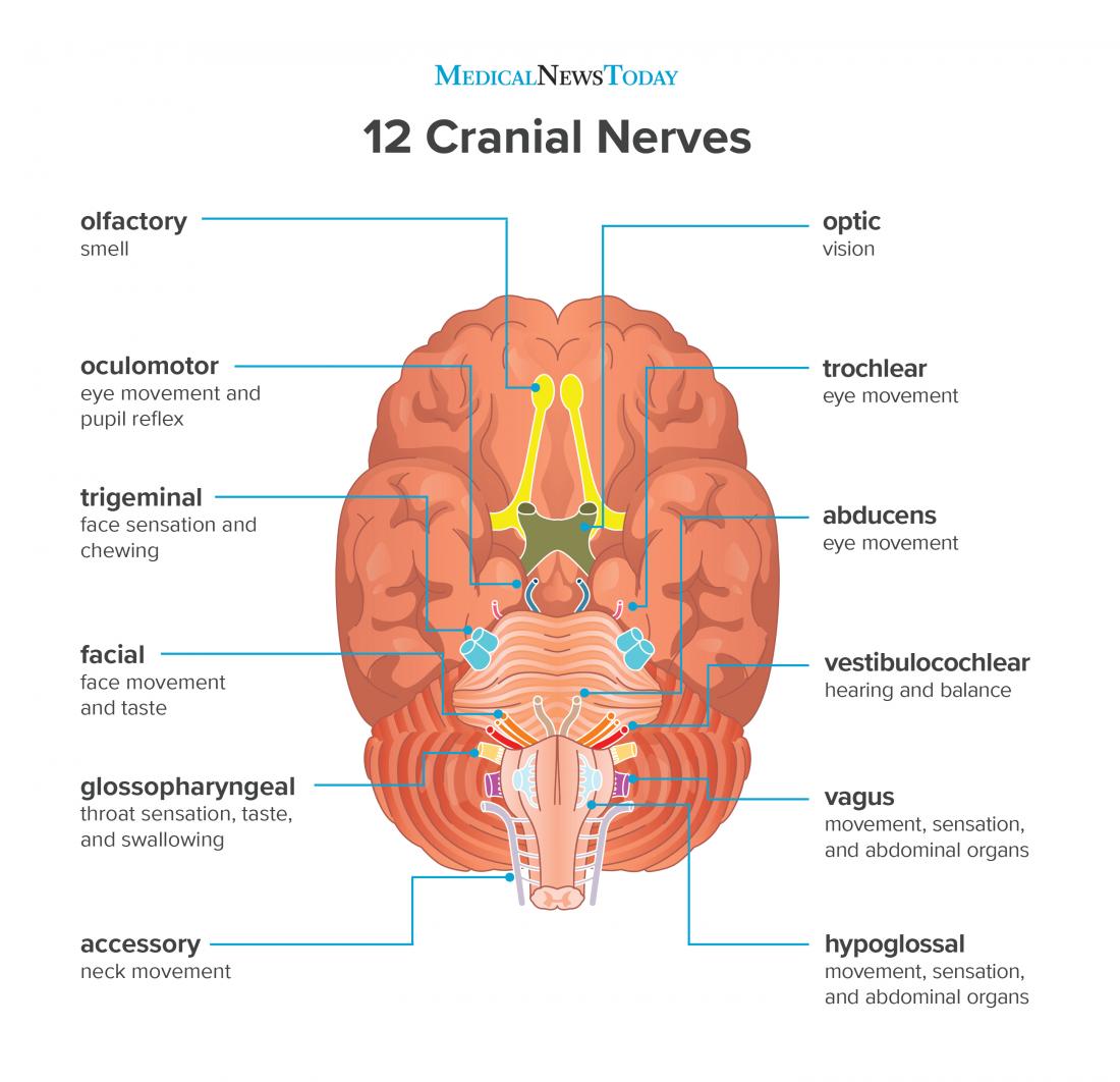
The cranial nerves are a set of twelve nerves that originate in the brain. Each has a different function for sense or movement.
The functions of the cranial nerves are sensory, motor, or both:
- Sensory cranial nerves help a person to see, smell, and hear.
- Motor cranial nerves help control muscle movements in the head and neck.
Each nerve has a name that reflects its function and a number according to its location in the brain. Scientists use Roman numerals from I–XII to label the cranial nerves in the brain.
This article will explore the functions of the cranial nerves and provide a diagram.
The olfactory nerve transmits information to the brain regarding a person’s sense of smell.
When a person inhales fragrant molecules, olfactory receptors within the nasal passage send the impulses to the cranial cavity, which then travel to the olfactory bulb.
Specialized olfactory neurons and nerve fibers meet with other nerves, which pass into the olfactory tract.
The olfactory tract then travels to the frontal lobe and other areas of the brain that are involved with memory and notation of different smells.
The
When light enters the eye, it hits the retina, which contains rods and cones. These are photoreceptors that translate signals from light into visual information for the brain.
Cones are located in the central retina and are involved with color vision. Rods are located in the peripheral retina and are involved with non-color vision.
These photoreceptors carry signal impulses along nerve cells to form the optic nerve. Most of the fibers of the optic nerve cross into a structure called the optic chiasm. Then, the optic tract projects to the primary visual cortex in the occipital lobe at the back of the brain. The occipital lobe is where the brain handles visual information.
The oculomotor nerve helps control muscle movements of the eyes.
The oculomotor nerve provides movement to most of the muscles that move the eyeball and upper eyelid, known as extraocular muscles.
The oculomotor nerve also helps with involuntary functions of the eye:
- The sphincter pupillae muscle automatically constricts the pupil to allow less light into the eye when the light is bright. When it is dark, the muscle relaxes to allow more light to enter.
- The ciliary muscles help the lens adjust to short range and long range vision. This happens automatically when a person looks at near or far objects.
The
The trochlear nerve, like the oculomotor nerve, originates in the midbrain. It powers the contralateral superior oblique muscle that allows the eye to point downward and inward.
The
Its motor functions help a person to chew and clench the teeth and gives sensation to muscles in the tympanic membrane of the ear.
Its sensory division has three parts that connect to sensory receptor sites on the face:
- The ophthalmic part gives sensation to parts of the eyes, including the cornea, mucosa in the nose, and skin on the nose, the eyelid, and the forehead.
- The maxillary part gives sensation to the middle third of the face, side of the nose, upper teeth, and lower eyelid.
- The mandibular part gives sensation to the lower third of the face, the tongue, mucosa in the mouth, and lower teeth.
Trigeminal neuralgia is a common disorder of the trigeminal nerve that can cause intense pain and facial tics.
The abducens nerve also helps control eye movements.
It helps the lateral rectus muscle, which is one of the extraocular muscles, to turn the gaze outward.
The abducens nerve starts in the pons of the brainstem, enters an area called Dorello’s canal, travels through the cavernous sinus, and ends at the lateral rectus muscle within the bony orbit.

The
The facial nerve is made up of four nuclei that serve different functions:
- movement of muscles that produce facial expression
- movement of the lacrimal, submaxillary, and submandibular glands
- the sensation of the external ear
- the sensation of taste
The four nuclei originate in the pons and medulla and join together to travel to the geniculate ganglion.
Bell’s palsy is a common disorder of the facial nerve, which causes paralysis on one side of the face and possibly loss of taste sensation.
The vestibulocochlear nerve is involved with a person’s hearing and balance.
The vestibulocochlear nerve contains two components:
- The vestibular nerve helps the body sense changes in the position of the head with regard to gravity. The body uses this information to maintain balance.
- The cochlear nerve helps with hearing. Specialized inner hair cells and the basilar membrane vibrate in response to sounds and determine the frequency and magnitude of the sound.
These fibers combine in the pons and exit the skull via the internal acoustic meatus in the temporal bone.
The
- The sensory function receives information from the throat, tonsils, middle ear, and back of the tongue. It is also involved with the sensation of taste for the back of the tongue.
- The motor division provides movement to the stylopharyngeus, which is a muscle that allows the throat to shorten and widen.
The glossopharyngeal nerve starts in the medulla oblongata in the brain and leaves the skull through the jugular foramen, which leads to the tympanic nerve.
The vagus nerve has a range of functions, providing motor, sensory, and parasympathetic functions.
- The sensory part provides sensation to the outer part of the ear, the throat, the heart, abdominal organs. It also plays a role in taste sensation.
- The motor part provides movement to the throat and soft palate.
- The parasympathetic function regulates heart rhythm and innervates the smooth muscles in the airway, lungs, and gastrointestinal tract.
The vagus nerve is the longest cranial nerve as it starts in the medulla and extends to the abdomen.
Doctors use vagus nerve stimulation therapy to treat various conditions, including epilepsy, depression, and anxiety. Learn more about the vagus nerve and stimulation therapy here.

The accessory nerve provides motor function to some muscles in the neck:
It controls the sternocleidomastoid and trapezius muscles that allow a person to rotate, extend, and flex the neck and shoulders.
The accessory nerve separates into spinal and cranial parts.
The spinal component starts in the spinal cord and travels into the skull through the foramen magnum. From there, it meets the cranial component of the accessory nerve and exits the skull along the internal carotid artery.
The cranial part of the accessory nerve combines with the vagus nerve.
The
The hypoglossal nerve originates in the medulla.
Disorders of the hypoglossal nerve can cause paralysis of the tongue, most often occurring on one side.
The twelve cranial nerves are a group of nerves that start in the brain and provide motor and sensory functions to the head and neck.
Each cranial nerve has its unique anatomical characteristics and functions.
Doctors can identify neurological or psychiatric disorders by testing cranial nerve functions.
0Comments