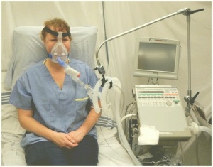
Hypercapnia is a common presentation in critically ill patients, with the potential for severe harm if not addressed appropriately. Hypoxaemia refers to a lower than normal arterial blood oxygen level, measured either as oxygen saturation (SaO2) or partial pressure of oxygen (PaO2).
- It is a common feature of acutely unwell hospitalised patients and can result in substantial morbidity and mortality if not treated rapidly and appropriately. Hypoxaemic patients may require admission to an intensive care unit, with more than 60% of those that do eventually requiring invasive ventilation.
- The mortality of hypoxaemic critically ill patients is 27%, rising to as high as 50% in patients with severe hypoxaemia No specific threshold of SaO2 or PaO2 defines hypoxaemia.
- Normal values for PaO2 are 10.5–13.5 kPa, and for SaO2 are 94–98%. Obtained via arterial blood gas (providing PaO2and SaO2) and pulse oximetry (providing peripheral capillary oxygen saturation; SpO2).
- Normal values decline with age and are influenced by the presence of comorbidities such as chronic lung disease.
Respiratory Failure: a clinical condition that happens when the respiratory system fails to maintain its main function, which is gas exchange, in which PaO2 is lower ie hypoxemia than 60 mmHg and/or PaCO2 higher than 50 mmHg. Respiratory failure is divided into type I and type II.
- Type I respiratory failure involves low oxygen, and normal or low carbon dioxide levels.
- Type II respiratory failure involves low oxygen, with high carbon dioxide.
Hypoxia and Hypoxema
The term hypoxia and hypoxemia are not synonymous.
- Hypoxemia is defined as a decrease in the partial pressure of oxygen in the blood
- Hypoxia is defined by reduced level of tissue oxygenation. Severe hypoxia can affect the production of ATP by mitochondrial oxidative phosphorylation, threatening cellular integrity. Non-oxygen dependent bioenergetic pathways are referred to as anaerobic metabolism; they are short-term inefficient systems that are unable to sustain life for prolonged periods of time in humans.
Mechanisms of Hypoxemia
There are various mechanisms of hypoxemia. These are V/Q mismatch, right-to-left shunt, diffusion impairment, hypoventilation, and low inspired PO2.
This is the most common type of hypoxemia. Ventilation refers to the oxygen supply in the lungs, while perfusion refers to the blood supply to the lungs.Ventilation/perfusion (V/Q) mismatch Image 2: Positive study pulmonary embolism situated within the right middle lobe/anterior segment of the right upper lobe and the posterior basal segment of the right lower lobe.
- Ventilation and perfusion are measured in a ratio, called V/Q ratio. Normally, there’s a small degree of mismatch in this ratio, however if the mismatch becomes too great, problems can occur.
- There are two causes of ventilation perfusion mismatch:
- The lungs are getting enough oxygen, but there’s not enough blood flow (increased V/Q ratio).
- There is blood flow to the lungs, but not enough oxygen (decreased V/Q ratio).
Shunt
- Normally, deoxygenated blood enters the right side of the heart, travels to the lungs to receive oxygen, and then travels to the left side of the heart to be distributed to the rest of the body.
- In this type of hypoxemia, blood enters the left side of the heart without becoming oxygenated in the lungs.
Diffusion impairment
- When oxygen enters the lungs, it fills the alveoli. Capillaries surround the alveoli. Oxygen diffuses from the alveoli into the blood running through the capillaries.
- In this type of hypoxemia, the diffusion of oxygen into the bloodstream is impaired.
Hypoventilation
- Hypoventilation is when oxygen intake occurs at a slow rate. This can result in higher levels of carbon dioxide in the blood and lower levels of oxygen.
Low Inspired PO2
- This type of hypoxemia typically occurs at higher altitudes. Available oxygen in the air decreases with increasing altitude.
- Therefore, at higher altitudes each breath provides you with lower levels of oxygen than when you’re at sea level.
Clinical Signs
A patient with acute hypoxaemia will display some or all of the following symptoms;
Chronic hypoxaemia can occur from chronic lung conditions such as COPD or seep apnoea, but it can also be caused by environmental factors such as frequent flying or living at high altitudes. which can be compensated or uncompensated. The compensation may result in the symptoms to be overlooked initially, however, further disease progression or mild illness such as a chest infection may increase oxygen demand and unmask the existing hypoxaemia.
Causes of Hypoxaemia
Some of the respiratory causes of hypoxaemia are:
- Anemia: a condition in which there aren’t enough red blood cells to effectively carry oxygen. Because of this, a person with anemia may have low levels of oxygen in their blood.
- Asthma
- Pulmonary embolus (PE): is a blockage of an artery in the lungs by a clot that has moved from elsewhere in the body through the bloodstream (embolism). Symptoms of a PE may include shortness of breath and chest pain, particularly on inspiration.
- Collapsed lung (atelectasis)
- Congenital heart defects or disease
- High altitudes
- Interstitial lung disease eg Pulmonary fibrosis: describes a collection of diseases which lead to interstitial lung damage and ultimately fibrosis and loss of the elasticity of the lungs. It is a chronic condition characterised by shortness of breath. The lung tissue becomes thickened, stiff and scarred over a period of time. The development of scar tissue is called fibrosis. As the lung tissue becomes scarred and thicker, the lungs start to lose their ability to transfer oxygen into the bloodstream.
- Medications that lower breathing rate eg some opiods and anesthetics
- Pneumonia: is caused by an infection which can start off as a lower respiratory tract infection, which when untreated can cause consolidation and significant sputum retention. Due to the lobe consolidation, the lungs are not adequately ventilated.
- Sleep apnea
- Pulmonary oedema: occurs when fluid accumulates in the alveoli of the lungs causing an increased work of breathing. This fluid accumulation interferes with gas exchange and can cause respiratory failure.
- Chronic respiratory conditions: such as COPD ( flow of air in the lungs is obstructed. Destruction of the walls of alveoli and surrounding capillaries in COPD can lead to problems with oxygen exchange, which can lead to hypoxemia) or CF
- Distended abdomen, e.g. pancreatitis, ascites. This prevents the diaphragm from descending which reduces the surface area for gas exchange.
- Hypoxemia can sometimes occur in newborns with congenital heart defects or disease. In fact, measuring the levels of oxygen in the blood is used to screen infants for congenital heart defects.
- Preterm infants are also vulnerable to hypoxemia, particularly if they’ve been placed on a mechanical ventilator
Medical Treatment of Hypoxaemia
The primary treatment for hypoxaemia is controlled oxygen therapy, alongside the identification and treatment of the underlying cause.
- Controlled Oxygen Therapy
Patients who are unable to maintain a SaO2 >90% with a face mask may require additional respiratory support. This might include either continuous positive airway pressure (CPAP), non-invasive ventilation or intubation with mechanical ventilation.
Controlled oxygen therapy is prescribed for hypoxaemic patients to increase alveolar oxygen tension and decrease work of breathing. It is important to remember that oxygen is a drug and should always be prescribed with the required percentage and/or flow rate. - Humidification (image 3)
Oxygen therapy is known to dry out the airways. Humidifying oxygen is often used in an attempt to help prevent the drying of the upper respiratory tract. In patients who are requiring high flow oxygen or oxygen for longer periods consider cold or heated humidification. Heated humidification is believed to be better for tenacious secretions or severe bronchospasm. - Treat the Cause
Whilst the delivery of oxygen therapy is the primary treatment for hypoxaemia it is essential to treat the cause ,e.g. bronchospasm, sputum retention, volume loss. There may be an acute primary respiratory problem, however, the respiratory failure could be due to compensation for another condition such as cardiac or renal. Multi-disciplinary team (MDT) working is vital in managing this.
Physiotherapy
The overall aim of physiotherapy is to identify and treat the cause of the hypoxaemia, thus aiming to increase PaO2 >8kPa while administering appropriate oxygen therapy. The cause of hypoxaemia may be sputum retention in which case various physiotherapy techniques can be used:
- Positioning - to improve V/Q matching
- Active cycle of breathing exercises
- Autogenic drainage
- Manual techniques: percussion or vibrations
- Bubble PEP or other oscillating instruments: acapella, flutter
- Cough assist or IPPB
- Home oxygen therapy
Complications with Hypoxaemia
If left untreated hypoxaemia can lead to type 1 respiratory failure. This may result is further symptoms which can be life threatening:
- Respiratory acidosis
- Cardiac arrhythmia
- Cerebral hypoxaemia
- Altered mental state including coma
- Cardiorespiratory arrest


0Comments