
Madelung deformity (MD) of the wrist is characterized by a growth disturbance in the volar-ulnar distal radial physis that results in a volar and ulnar tilted distal radial articular surface, volar translation of the hand and wrist, and a dorsally prominent distal ulna. It occurs predominantly in adolescent females who present with pain, decreased range of motion (ROM), and deformity. MD often has a genetic etiology and is associated with mesomelic dwarfism and a mutation on the X chromosome.

Madelung Deformity
The deformity can be treated surgically by addressing the deforming bony and ligamentous lesions, correcting the abnormal position of the radial articular surface, and equalizing the longitudinal levels of the distal radius and ulna.
At the Seventh German Surgical Congress of 1878, Otto W Madelung described the wrist deformity bearing his name as "Die spontane Subluxation der Hand nach vorne" ("spontaneous forward subluxation of the hand"). Although previous authors had described lesions that we might now consider examples of MD, the ascribed etiologies were inconsistent.
Several authors before Madelung had described various entities termed carpus curvus, radius curvus, progressive subluxation of the wrist, manus valga, manus furca, and idiopathic progressive curvature of the radius. Dupuytren's 1834 description was based on a quotation from Bégin, who attributed the deformity to a repetitive occupational injury. In 1847, Nelaton vaguely described an anatomic specimen with a deformity suggestive of MD. In 1855, Malgaigne also described a good example of MD. However, it was Madelung who first provided an accurate clinical description and proposed both an etiology and treatment.
Contributions to the French, German, and Italian literature concerning MD between 1880 and 1908 were purely descriptive, without agreement regarding etiology, pathogenesis, or treatment. During the same period, however, Madelung's work in the British and American literature was completely disregarded. One of the first papers on this topic in the American literature, a case report and review of the European literature authored by DeWitt Stetten in 1909, only perpetuated inaccurate information.
In 1938, Anton, Reitz, and Spiegel published the first comprehensive paper on MD in the American literature in the Annals of Surgery. They collected and tabulated 172 cases of MD and gave a detailed case history of one case. They also presented clinical, radiographic, pathologic, epidemiologic, and etiologic theories regarding MD. Anton et al attempted to create a new classification system and even to rename the deformity from its eponym to a more descriptive term; however, their main purpose was to dispel the inaccurate information presented in previous reports.
Madelung deformity (MD) of the wrist is characterized by a growth disturbance in the volar-ulnar distal radial physis that results in a volar and ulnar tilted distal radial articular surface, volar translation of the hand and wrist, and a dorsally prominent distal ulna. It occurs predominantly in adolescent females who present with pain, decreased range of motion (ROM), and deformity. MD often has a genetic etiology and is associated with mesomelic dwarfism and a mutation on the X chromosome.
 |
| Madelung Deformity |
The deformity can be treated surgically by addressing the deforming bony and ligamentous lesions, correcting the abnormal position of the radial articular surface, and equalizing the longitudinal levels of the distal radius and ulna.
At the Seventh German Surgical Congress of 1878, Otto W Madelung described the wrist deformity bearing his name as "Die spontane Subluxation der Hand nach vorne" ("spontaneous forward subluxation of the hand"). Although previous authors had described lesions that we might now consider examples of MD, the ascribed etiologies were inconsistent.
Several authors before Madelung had described various entities termed carpus curvus, radius curvus, progressive subluxation of the wrist, manus valga, manus furca, and idiopathic progressive curvature of the radius. Dupuytren's 1834 description was based on a quotation from Bégin, who attributed the deformity to a repetitive occupational injury. In 1847, Nelaton vaguely described an anatomic specimen with a deformity suggestive of MD. In 1855, Malgaigne also described a good example of MD. However, it was Madelung who first provided an accurate clinical description and proposed both an etiology and treatment.
Contributions to the French, German, and Italian literature concerning MD between 1880 and 1908 were purely descriptive, without agreement regarding etiology, pathogenesis, or treatment. During the same period, however, Madelung's work in the British and American literature was completely disregarded. One of the first papers on this topic in the American literature, a case report and review of the European literature authored by DeWitt Stetten in 1909, only perpetuated inaccurate information.
In 1938, Anton, Reitz, and Spiegel published the first comprehensive paper on MD in the American literature in the Annals of Surgery. They collected and tabulated 172 cases of MD and gave a detailed case history of one case. They also presented clinical, radiographic, pathologic, epidemiologic, and etiologic theories regarding MD. Anton et al attempted to create a new classification system and even to rename the deformity from its eponym to a more descriptive term; however, their main purpose was to dispel the inaccurate information presented in previous reports.
Anatomy
An important anatomic consideration in MD is the normal position of the distal radial articular surface. The following four features of this articular surface should be noticed radiographically:
• Radial inclination - This is the angle formed by a line from the distal radioulnar joint (DRUJ) to the radial styloid and a line perpendicular to the shaft of the radius through the lunate fossa; it is normally 21-23°
• Radial length - This is the difference in longitudinal level between the lunate fossa and the radial styloid; it is normally 12-15 mm
• Volar tilt - This is measured on a lateral radiograph and is the angle formed between a line perpendicular to the radial shaft and a line through the dorsal and volar rims of the radial joint surface; normally, it is 10-15°
• Ulnar variance - This is the relative difference in height between the radial and ulnar distal articular surfaces; these should be level with each other
An important anatomic consideration in MD is the normal position of the distal radial articular surface. The following four features of this articular surface should be noticed radiographically:
• Radial inclination - This is the angle formed by a line from the distal radioulnar joint (DRUJ) to the radial styloid and a line perpendicular to the shaft of the radius through the lunate fossa; it is normally 21-23°• Radial length - This is the difference in longitudinal level between the lunate fossa and the radial styloid; it is normally 12-15 mm
• Volar tilt - This is measured on a lateral radiograph and is the angle formed between a line perpendicular to the radial shaft and a line through the dorsal and volar rims of the radial joint surface; normally, it is 10-15°
• Ulnar variance - This is the relative difference in height between the radial and ulnar distal articular surfaces; these should be level with each other
Pathophysiology
One third of cases of MD are transmitted in an autosomal dominant fashion. The condition has a variable expression and 50% penetrance. MD is bilateral in 50% of cases and is primarily found in females. A number of affected kindreds of patients with MD have been described. Numerous cases of multiple patients with MD within families have been reported. One report detailed five generations of family members with bilateral MD without signs of dyschondrosteosis.
Finally, a primary chromosomal association with MD has been observed in patients with Turner syndrome (karyotype XO). MD was the presenting sign of Turner syndrome in one young girl, while Henry and Thorburn diagnosed Turner syndrome incidentally when studying a series of patients with MD who underwent cytogenetic evaluation.
Molecular genetic studies clarified the association of the female predominant MD, dyschondrosteosis, and the missing X chromosome in Turner syndrome. The idea that an X chromosomal translocation caused dyschondrosteosis was first proposed in 1985. An X chromosomal translocation also was found to be associated with MD in 1997.
In 1998, groups from London, England, and Switzerland established a marker that was linked to the pseudoautosomal region (PAR1) of the X and Y chromosomes. Within families affected by a short stature dysplasia, the groups found deletions and a premature stop codon (exon 4) in the short stature homeobox-containing gene, SHOX, that segregated the marker in band Xp22.
In 2000, another group reported that the SHOX gene mutation was found in patients with dyschondrosteosis and MD in multiple cases. Families with this mutation and individuals with Turner syndrome (both essentially hemizygous individuals for the SHOX gene) and families with a history of MD have been shown to exhibit a variable expression of MD and dyschondrosteosis. This variability indicates that a modifier gene on another area of the X chromosome or on an autosomal gene most likely is involved also. The interaction of the SHOX locus with other genes and transcription factors is not known. The SHOX mutation continues to be the subject of many studies.
Etiology
Henry and Thorburn classified MD into the following four etiologic groups [9] :
• Posttraumatic
• Dysplastic
• Chromosomal or genetic (Turner syndrome)
• Idiopathic or primary
Posttraumatic MD has been found following repetitive trauma or following a single event that disrupts growth of the distal radial ulnar-volar physis. Bone dysplasias associated with MD include the following:
• Multiple hereditary osteochondromatosis
• Ollier disease
• Achondroplasia
• Multiple epiphyseal dysplasias
• Mucopolysaccharidoses (eg, Hurler and Morquio syndromes)
Secondary causes of wrist deformity that may mimic MD include the following:
• Sickle-cell disease
• Infection
• Tumor
• Rickets
The most important dysplasia associated with MD, however, is dyschondrosteosis.
Dyschondrosteosis is a form of mesomelic dwarfism associated with MD that was first described by Leri and Weill in 1929. It is characterized by variable short stature, short forearms, and tibial/fibular shortening. Specifically, height is less than the 25th percentile, the radius is 75% of the length of the humerus, and the tibia is 85% of the length of the femur. Dyschondrosteosis becomes more clinically pronounced during adolescence. No other abnormalities are commonly associated with it.
Forearm shortening in dyschondrosteosis tends to be bilateral and appears almost identical to that in primary MD, except that the proximal radius is involved in patients with dyschondrosteosis (see the first image below), and the proximal radius is not involved in primary MD (see the second and third images below). Both dyschondrosteosis and MD are transmitted in an autosomal dominant fashion with a female predominance.
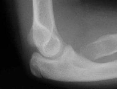
Lateral radiograph of elbow of patient A, depicting a dysplastic proximal radius. This is characteristic of dyschondrosteosis.
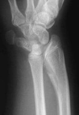
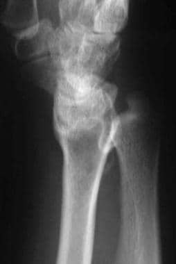
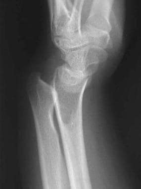
Preoperative anteroposterior radiograph of wrist of patient B. This patient has primary Madelung deformity (no sign of dyschondrosteosis).
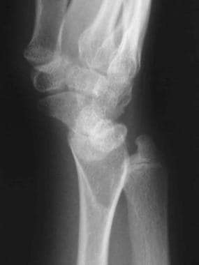
Preoperative lateral radiograph of wrist of patient B. The flame-shaped radiolucency in the metaphysis of the radius is occupied by the fibrocartilaginous Vickers ligament.
With such a similarity between primary MD and Leri-Weill dyschondrosteosis, the reason why many authors have described them as a single entity is apparent. However, many children with unilateral and bilateral MD have normal stature and no other characteristic of dyschondrosteosis. Therefore, it is reasonable to describe primary MD as a separate, albeit related, condition.
To differentiate primary MD from dyschondrosteosis, Felman and Kirkpatrick suggested the following criteria : Primary MD can be defined as occurring in a child above the 25th percentile in height with no family history of dyschondrosteosis and no history of other secondary causes. Conversely, a patient who is shorter than 5 ft (~1.5 m) at skeletal maturity, has proximal involvement of the radius, and has a relatively short tibia and fibula may have mesomelic dwarfism.
Henry and Thorburn classified MD into the following four etiologic groups [9] :
• Posttraumatic• Dysplastic
• Chromosomal or genetic (Turner syndrome)
• Idiopathic or primary
Posttraumatic MD has been found following repetitive trauma or following a single event that disrupts growth of the distal radial ulnar-volar physis. Bone dysplasias associated with MD include the following:
• Multiple hereditary osteochondromatosis• Ollier disease
• Achondroplasia
• Multiple epiphyseal dysplasias
• Mucopolysaccharidoses (eg, Hurler and Morquio syndromes)
Secondary causes of wrist deformity that may mimic MD include the following:
• Sickle-cell disease• Infection
• Tumor
• Rickets
The most important dysplasia associated with MD, however, is dyschondrosteosis.
Dyschondrosteosis is a form of mesomelic dwarfism associated with MD that was first described by Leri and Weill in 1929. It is characterized by variable short stature, short forearms, and tibial/fibular shortening. Specifically, height is less than the 25th percentile, the radius is 75% of the length of the humerus, and the tibia is 85% of the length of the femur. Dyschondrosteosis becomes more clinically pronounced during adolescence. No other abnormalities are commonly associated with it.
Forearm shortening in dyschondrosteosis tends to be bilateral and appears almost identical to that in primary MD, except that the proximal radius is involved in patients with dyschondrosteosis (see the first image below), and the proximal radius is not involved in primary MD (see the second and third images below). Both dyschondrosteosis and MD are transmitted in an autosomal dominant fashion with a female predominance.
|
| Lateral radiograph of elbow of patient A, depicting a dysplastic proximal radius. This is characteristic of dyschondrosteosis. |
|
| Preoperative anteroposterior radiograph of wrist of patient B. This patient has primary Madelung deformity (no sign of dyschondrosteosis). |
|
| Preoperative lateral radiograph of wrist of patient B. The flame-shaped radiolucency in the metaphysis of the radius is occupied by the fibrocartilaginous Vickers ligament. |
With such a similarity between primary MD and Leri-Weill dyschondrosteosis, the reason why many authors have described them as a single entity is apparent. However, many children with unilateral and bilateral MD have normal stature and no other characteristic of dyschondrosteosis. Therefore, it is reasonable to describe primary MD as a separate, albeit related, condition.
To differentiate primary MD from dyschondrosteosis, Felman and Kirkpatrick suggested the following criteria : Primary MD can be defined as occurring in a child above the 25th percentile in height with no family history of dyschondrosteosis and no history of other secondary causes. Conversely, a patient who is shorter than 5 ft (~1.5 m) at skeletal maturity, has proximal involvement of the radius, and has a relatively short tibia and fibula may have mesomelic dwarfism.
Approach Considerations
Operative treatment of Madelung deformity (MD) is indicated for pain relief and cosmetic improvement. These indications are consistent among many authors. Range of motion (ROM), especially in pronation and supination, usually is only minimally improved and has varied in different operative series.
In the skeletally immature patient with clear evidence of MD, the most likely cause of pain is tension within the Vickers ligament. Release of the ligament either alone or in combination with an osteotomy is indicated.
In the skeletally mature patient, the congruency of the radiocarpal joint and that of the distal radioulnar joint (DRUJ) are assessed. If an osteotomy will result in a secondarily congruous joint, then an osteotomy is indicated. However, if radiocarpal congruity is not possible to obtain, a radioscaphocapitate arthrodesis is indicated. Similarly, if DRUJ congruity is not possible, then a Darrach or Sauve-Kapandji procedure is indicated.
No specific contraindications for surgery exist other than those associated with any elective surgical procedure.
Nonoperative Therapy
Nonoperative management may be helpful in skeletally mature individuals with MD and mild-to-moderate short-term wrist pain. If the pain is predominantly related to the DRUJ, then a sugar tong–type splint may relieve joint irritation from overactivity. If radiocarpal pain is the primary problem, then a volar splint may decrease symptoms. Patients may eventually decrease their manual activity levels to a point where their symptoms are manageable without surgery.
In contrast, the younger and skeletally immature patient with clear evidence of MD has pain that is caused by the tension within the Vickers ligament, and splinting most likely will not have a satisfactory result.
Surgical Options
The surgical decision is based on the following four factors:
• Patient's age and the growth remaining in the distal radius• Severity of the deformity
• Severity of the symptoms
• Clinical and radiographic findings
Operative treatment can be divided into three broad categories as follows:
• Procedures that correct the primary deformity of the radius• Procedures that attempt to decrease pain and increase ROM by making a compensatory change in the ulna
• Procedures that address both
Multiple procedures address the deformity in the radius. Conceptually, these can be broken into the following three types:
• Procedures that change the growth or anatomy at the physis• Procedures that change the bony anatomy of the metaphysis
• Procedures that are considered salvage-type joint-sacrificing procedures
The goal of ulnar procedures is to change the relationship of the relatively long ulna to the radius.
Vickers Physiolysis
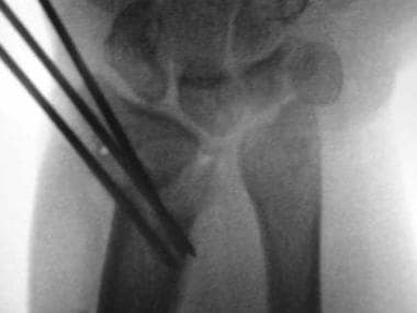
When the deformity is noticed early and significant growth remains, changing the growth pattern of the distal radial physis to correct the deformity is possible. In 1992, Vickers and Nielsen described the lesion in the volar and ulnar distal radius as both bony and ligamentous, and they stated that it is an inherent failure of focal growth and structural tethering of further growth. They described an ulnar-volar release for MD of the physis, called physiolysis (see the images below).
 |
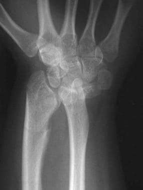
| Postoperative anteroposterior radiograph of wrist of patient B following Vickers physiolysis. Vickers ligament and the ulnar abnormal physis have been excised. |
This allows, then, normal and compensatory growth to correct the deformity, much like the bony bridge resection of Langenskiöld. Vickers and Nielsen used the procedure of resecting the bony and ligamentous lesion (the first to correct MD using remaining growth to correct the deformity). They were the first to describe a ligamentous lesion as part of the pathology and also were the first to use the volar approach to address it.
Through a volar flexor carpi radialis (FCR) approach, the pronator is mobilized, and the distal radius is exposed. An osteotome is used to make a longitudinal split 5 mm from the DRUJ on the radius, and parallel transverse 1-mm sections are made until normal physis is identified.
Some physis is left to overhang the metaphysis. Fat harvested from the forearm is packed into the bone defect. Completely excising the tethering ligament (Vickers ligament) between the lunate and the radius is critical. (See the images below.)
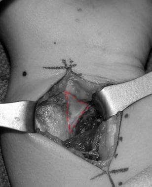 |
| Intraoperative photo of Vickers ligament, outlined in red. |
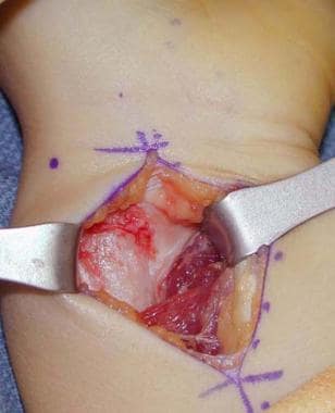 |
| Intraoperative color photograph of Vickers ligament. The ligament is outlined in the previous image. |
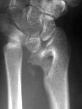
The Vickers ligament originates on the radius in a fossa that is seen radiographically as a flame-shaped radiolucency distal to a bone spur on the ulnar aspect of the distal metaphysis (see the images below). Dannenberg noted this characteristic as an area of osteopenia in his original radiographic criteria in 1938. Carter and Ezaki noted it in 91% of their cases.
 |
| Preoperative posteroanterior radiograph of wrist from patient C. |
 |
| Preoperative lateral radiograph of wrist from patient C. |
The Vickers ligament is fibrous and fibrocartilaginous and 5-7 mm thick. It inserts into the anterior surface of the lunate and the anterior radioulnar ligament portion of the triangular fibrocartilage complex (TFCC). Most likely, it is a secondary pathologic structure to a primary bony derangement and may even be a coalescence of normal structures.
This ligament may be an etiologic factor in MD pain. Following operative release of this ligament, patients state soon after surgery that their pain is largely relieved.
Radial Osteotomy
If the deformity has progressed in an older child and remaining growth is insufficient, several procedures can be used to correct the position of the distal radiocarpal joint surface.
They generally consist of a biplane osteotomy, either closing or opening wedge, which corrects the position of the joint surface and brings the radius and ulna into a more proper position. If a positive ulnar variance remains, an ulna-shortening procedure can be performed.
A less commonly used method is distraction histogenesis with the Ilizarov technique. Several authors have described experience with the technique of radial osteotomy and subsequent angular and length correction over time. The goal is slow correction of the distal radius to make a more congruous DRUJ. Children treated with this method are either skeletally mature or nearly mature.
Half pins are commonly selected over tensioned thin wires to allow better ROM during distraction. Either three or four rings may be used, with fewer complications and less pain noted when three are used. The first goal after fixation placement and radial osteotomy is angular correction over a 2-week period. The second goal is lengthening of approximately 1.5 cm over the next 2 weeks, followed by consolidation for 3 weeks. The entire process in the distractor takes 8 weeks.
In 2000, Carter and Ezaki reported a combined procedure using a Vickers ligament release and a dome-shaped osteotomy of the radius to correct all of the aspects of the radial deformity, including the radial and volar translation of the distal metaphysis.
This procedure is performed with a volar approach, and the Vickers ligament is excised entirely.
A dome-shaped osteotomy is made in the metaphysis so that the articular surface not only decreases in its radial inclination but also translates as it rotates. This translation reestablishes articular support to the lunate.
A second maneuver translates the articular surface dorsally. The prominent volar bone is then removed, and the osteotomy is pinned into position with two Steinman pins. This not only corrects the deformity but also decreases pain and increases ROM. (See the images below.)
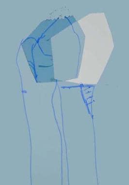 |
| Preoperative plan prior to Carter-Ezaki dome osteotomy. Dorsal translation of the distal radius is depicted. |
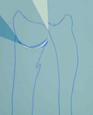 |
| Preoperative plan prior to Carter-Ezaki dome osteotomy. Rotation of the distal radius to accomplish both ulnar translation and normalization of radial tilt is depicted. |
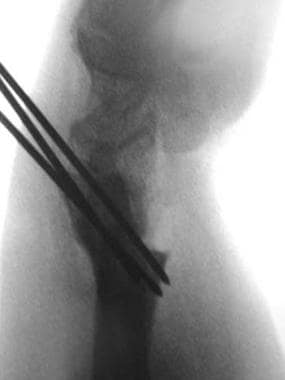 |
| Postoperative lateral radiograph. Note dorsal translation of distal radius after Carter-Ezaki dome osteotomy. |
 |
| Postoperative posteroanterior radiograph with Kirschner-wire fixation in place. Note combination of ulnar translation of distal radius and correction of radial tilt towards normal after Carter-Ezaki dome osteotomy. |
Volar ligament release with distal radial dome osteotomy has been shown to yield lasting correction of Madelung deformity.
Radioulnar Length Adjustment
In MD, the ulna grows normally and becomes longer than the radius. Because the radius is volar, the ulna appears to be subluxated dorsally. The incongruence at the DRUJ and the impingement of the radius on the ulna in supination may cause pain and contribute to decreased ROM in supination.
To allow unrestricted rotation, several ulnar procedures have been described; these include ulnar shortening, ulnar head resection and a Sauve-Kapandji (Lauenstein) DRUJ arthrodesis, [34] and ulnar pseudoarthrosis. Several authors have advocated both radial and ulnar procedures.
The Darrach procedure has long been a treatment option for MD. In isolation, this construct may leave the carpus unstable, especially in light of the increased ulnar and volar slope of the radial articular surface, as a result of which the carpus tends to slide off of the ulnar side of the wrist. Several authors have devised procedures to solve this problem.
The Sauve-Kapandji (Lauenstein) procedure may be a viable option for MD in that when the ulnar head is preserved, less chance exists for ulnar migration of carpus.
In 1975, Ranawat studied 13 wrists in eight patients with MD. His operative indications were pain and limitation of motion; deformity alone was not an indication for operation.
Mild deformity was treated with a Darrach procedure, and severe deformity was treated with Darrach plus a biplane radial osteotomy. In patients treated with a Darrach procedure, the carpus tended to translate ulnarward.
A radial osteotomy was theorized to improve the muscle balance about the wrist and to provide better axial bone support for the carpus on the radius in the absence of the ulnar head.
In 1993, Watson et al described another combined procedure in which the radial osteotomy was performed with both a closing wedge technique on the radial aspect of the metaphysis and an opening wedge on the ulnar aspect with the radial bone wedge.
This technique preserved radial length but also required an ulnar head resection. Pain was significantly relieved in all 15 wrists.
White and Weiland described a combination procedure in a series of wrists with MD of posttraumatic etiology.
The procedure included a volar closing wedge osteotomy along with a distal radioulnar arthrodesis and ulnar pseudoarthrosis proximal to the arthrodesis. The osteotomy of the radius improved the mechanics of the radiocarpal joint and prevented radiocarpal arthritis.
This procedure alone may not improve pain on supination/pronation due to the incongruent DRUJ.
Resection of the distal ulna, as in the Darrach procedure, may solve this problem, but ulnar translation of the carpus may result.
Fusing the DRUJ maintained carpal stability. Increase in the patient's ROM was minimal in any plane, but pain was significantly relieved, the carpus was stable, and the cosmetic result was satisfactory.
In the skeletally mature patient who presents with pain and in whom physical examination demonstrates limitation of motion and severe radiocarpal joint incongruity, a salvage procedure may be indicated.
When incongruity is severe, any surgical manipulation of the joint surfaces would be difficult, and the joint would not be stable for smooth articulation. For long-term pain relief and a stable joint, a radiocarpal arthrodesis and distal ulna resection are indicated.
Postoperative Care
Postoperative management depends on what is done operatively. Cast immobilization is necessary for 6-8 weeks after an osteotomy of the radius or ulna to allow for bony healing. Hand therapy is necessary in children who are not able to regain ROM on their own after 2-3 months without restrictions.
Complications
In the author's experience, no specific complications after operative correction have arisen as problematic. However, isolated cases of postoperative wound infections, reflex sympathetic dystrophy, and recurrent deformity after continued growth are reported in the literature.
Long-Term Monitoring
Children treated for MD should be monitored until surgical issues are resolved and then yearly until skeletal maturity is reached. After a physiolysis procedure, yearly radiographs can be used to document improvement in the position of the distal radial articular surface with growth.
0Comments