
A chest tube can help drain air, blood, or fluid from the space surrounding your lungs, called the pleural space.
Chest tube insertion is also referred to as chest tube thoracostomy. It’s typically an emergency procedure. It may also be done after surgery on organs or tissues in your chest cavity.
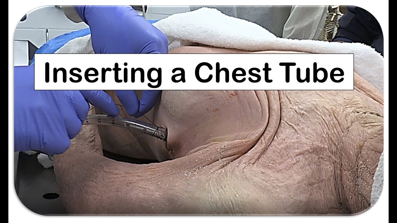 |
| Chest Tube Insertion |
During chest tube insertion, a hollow plastic tube is inserted between your ribs into the pleural space. The tube may be connected to a machine to help with the drainage. The tube will stay in place until the fluid, blood, or air is drained from your chest.
You may need a chest tube if you have any of the following:
- a collapsed lung
- a lung infection
- bleeding around your lung, especially after a trauma (such as a car accident)
- fluid buildup due to another medical condition, such as cancer or pneumonia
- breathing difficulty due to a buildup of fluid or air
- surgery, especially lung, heart, or esophageal surgery
Inserting a chest tube may also help your doctor diagnose other conditions, such as lung damage or internal injuries after a trauma.
Chest tube insertion is most commonly performed after surgery or as an emergency procedure, so there’s usually no way for you to prepare for it. Your doctor will ask for your consent to perform the procedure if you’re conscious. If you’re unconscious, they’ll explain why a chest tube was necessary after you wake up.
In cases where it isn’t an emergency, your doctor will order a chest X-ray before chest tube insertion. This is done to help confirm whether fluid or air buildup is causing the problem and to determine if a chest tube is needed. Some other tests may also be done to evaluate pleural fluid, such as a chest ultrasound or chest CT scan
Someone who specializes in lung conditions and diseases is called a pulmonary specialist. A surgeon or pulmonary specialist will usually perform the chest tube insertion. During chest tube insertion, the following happens:
Preparation: Your doctor will prepare a large area on the side of your chest, from your armpit down to your abdomen and across to your nipple. Preparation involves sterilizing the area and shaving any hair from the insertion site, if necessary. Your doctor may use an ultrasound to identify a good location for inserting the tube.
Anesthesia: The doctor may inject an anesthetic into your skin or vein to numb the area. The medication will help make you more comfortable during the chest tube insertion, which can be painful. If you’re having major heart or lung surgery, you’ll likely be given general anesthesia and be put to sleep before the chest tube is inserted.
Incision: Using a scalpel, your doctor will make a small (¼- to 1 ½-inch) incision between your ribs, near the upper part of your chest. Where they make the incision depends on the reason for the chest tube.
Insertion: Your doctor will then gently open a space into your chest cavity and guide the tube into your chest. Chest tubes come in various sizes for different conditions. Your doctor will stitch the chest tube in place to prevent it from moving. A sterile bandage will be applied over the insertion site.
Drainage: The tube is then attached to a special one-way drainage system that allows air or fluid to flow out only. This prevents the fluid or air from flowing back into the chest cavity. While the chest tube is in, you’ll probably need to stay in the hospital. A doctor or nurse will monitor your breathing and check for possible air leaks.
How long the chest tube is left in depends on the condition that caused the buildup of air or fluid. Some lung cancers can cause fluid to reaccumulate. Doctors may leave the tubes in for a longer period of time in these cases.
Chest tube insertion puts you at risk of several complications. These include:
Pain during placement: Chest tube insertion is usually very painful. Your doctor will help manage your pain by injecting an anesthetic through an IV or directly into the chest tube site. You’ll be given either general anesthesia, which puts you to sleep, or local anesthesia, which numbs the area.
Infection: As with any invasive procedure, there’s a risk of infection. The use of sterile tools during the procedure helps reduce this risk.
Bleeding: A very small amount of bleeding can occur if a blood vessel is damaged when the chest tube is inserted.
Poor tube placement: In some cases, the chest tube can be placed too far inside or not far enough inside the pleural space. The tube may also fall out.
Serious complications
Serious complications are rare, but they can include:
- bleeding into the pleural space
- injury to the lung, diaphragm, or stomach
- collapsed lung during tube removal
The chest tube usually stays in for a few days. After your doctor is sure that no more fluid or air needs to be drained, the chest tube will be removed.
The removal of the chest tube is usually performed quickly and without sedation. Your doctor will give you specific instructions on how to breathe when the tube is removed. In most cases, the chest tube will be removed as you’re holding your breath. This ensures extra air doesn’t get into your lungs.
After the doctor removes the chest tube, they’ll apply a bandage over the insertion site. You may have a small scar. Your doctor will likely schedule an X-ray at a later date to make sure that there isn’t another buildup of air or fluid inside your chest.
0Comments