
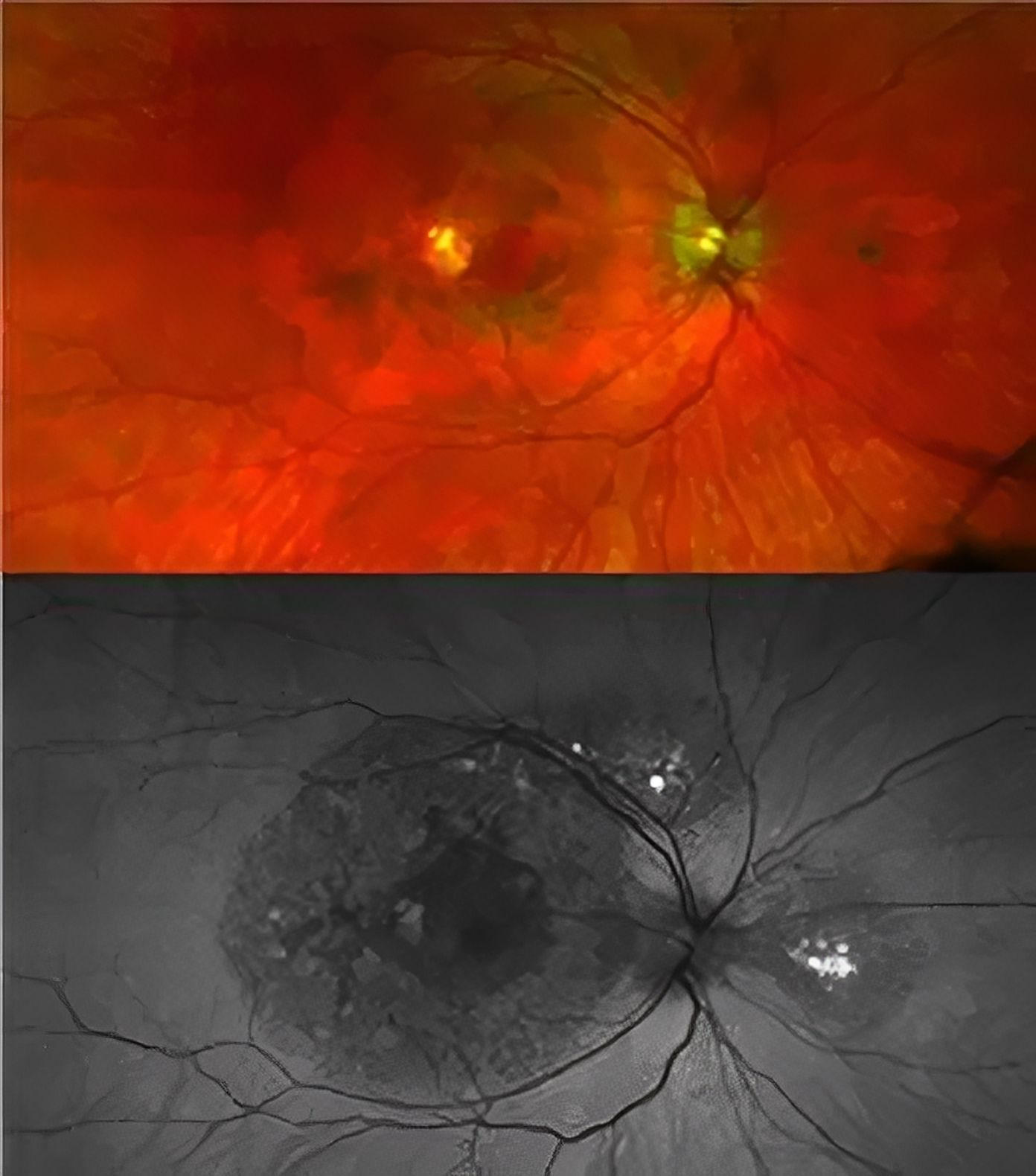
Retinal images of a patient with a TIMP3 mutation causing atypical symptoms. While there is visible damage in the retina (dark circles), there is no choroidal neovascularization present. Credit: National Eye Institute
A new type of macular dystrophy, which is a cause of central vision loss, has been discovered through genetic and clinical research.
A new disease that damages the macula, a small region of the light-sensing retina required for sharp, central vision, has been discovered by National Eye Institute (NEI) researchers. The researchers have published their findings on the unnamed new macular dystrophy in the journal JAMA Ophthalmology. NEI is a branch of the National Institutes of Health.
Macular dystrophies are disorders that often result in central vision loss due to abnormalities in various genes, including ABCA4, BEST1, PRPH2, and TIMP3.
For instance, individuals with Sorsby Fundus Dystrophy, a hereditary eye disorder that is specifically linked with TIMP3 variations, often develop symptoms in adulthood. Due to choroidal neovascularization, which is the growth of new, irregular blood vessels behind the retina that leak fluid and disrupt vision, they often experience abrupt changes in visual acuity.
TIMP3 is a protein that helps regulate retinal blood flow and is secreted from the retinal pigment epithelium (RPE), a layer of tissue that nourishes and supports the retina’s light-sensing photoreceptors. All TIMP3 gene mutations reported are in the mature protein after it has been “cut” from RPE cells in a process called cleavage.
“We found it surprising that two patients had TIMP3 variants not in the mature protein, but in the short signal sequence the gene uses to ‘cut’ the protein from the cells. We showed these variants prevent cleavage, causing the protein to be stuck in the cell, likely leading to retinal pigment epithelium toxicity,” said Bin Guan, Ph.D., lead author.
The research team followed these findings with clinical evaluations and genetic testing of family members to verify that the two new TIMP3 variants are connected to this atypical maculopathy.
“Affected individuals had scotomas, or blind spots, and changes in their maculas indicative of disease, but, for now, they have preserved central vision and no choroidal neovascularization, unlike typical Sorsby Fundus Dystrophy”, said Cathy Cukras, M.D., Ph.D., a Lasker tenure-track investigator and medical retina specialist who clinically evaluated the patients.
NEI’s Ophthalmic Genomics Laboratory gathers and manages specimens and diagnostic data from patients who have been recruited into multiple studies within the NEI clinical program to facilitate research of rare eye diseases, including Sorsby Fundus Dystrophy.
“Discovering novel disease mechanisms, even in known genes like TIMP3, may help patients that have been looking for the correct diagnosis, and will hopefully lead to new therapies for them,” said Rob Hufnagel, M.D., Ph.D., senior author, and director of the Ophthalmic Genomics Laboratory at NEI.
Reference: “Early-Onset TIMP3-Related Retinopathy Associated With Impaired Signal Peptide” by Bin Guan, Ph.D., Laryssa A. Huryn, MD, Andrew B. Hughes, BS, Zhiyu Li, MD, Chelsea Bender, BS, Delphine Blain, MS, MBA, Amy Turriff, MS, Catherine A. Cukras, MD, Ph.D. and Robert B. Hufnagel, MD, Ph.D., 9 June 2022, JAMA Ophthalmology.
DOI: 10.1001/jamaophthalmol.2022.1822
The study was funded by the NEI Intramural Research Program.
0Comments